Our approach at Lanterne Dx combines whole-slide imaging at single-cell resolution to visualize and quantitate biomarker expression. This reveals how cells interact and are organized across the entire tissue landscape.
Visualize up to 6 tissue protein biomarkers or 4 RNA biomarkers simultaneously in whole tissue slides with a multiplex panel. We can offer purchased off-the shelf ready to go panels, as well as develop fully customized panels. Your assays can be developed with ease and to meet your specific needs. Identify and quantify overlapping biomarkers for greater understanding of the tumor microenvironment leading to enhanced decisions for patient enrollment and accelerated development.
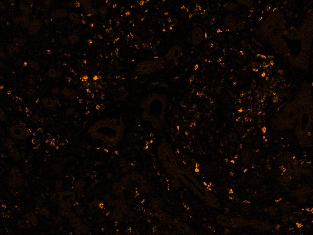
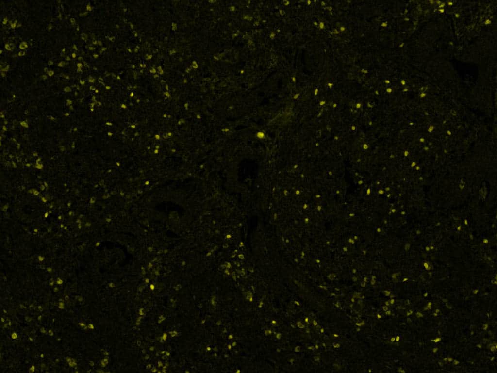
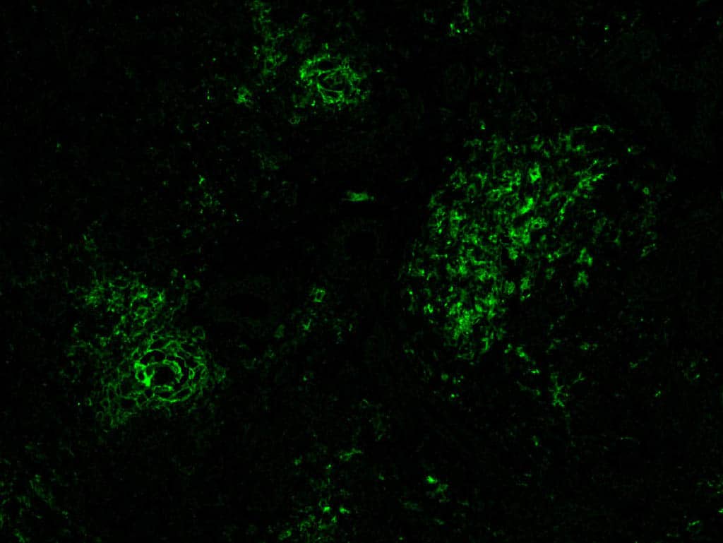
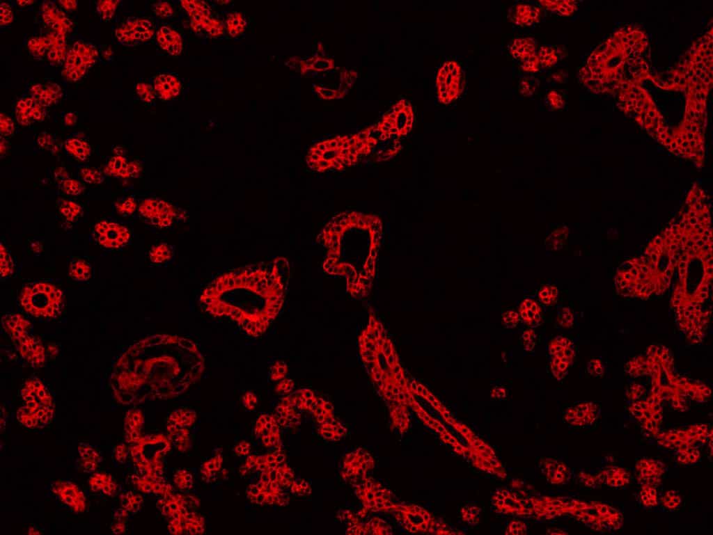
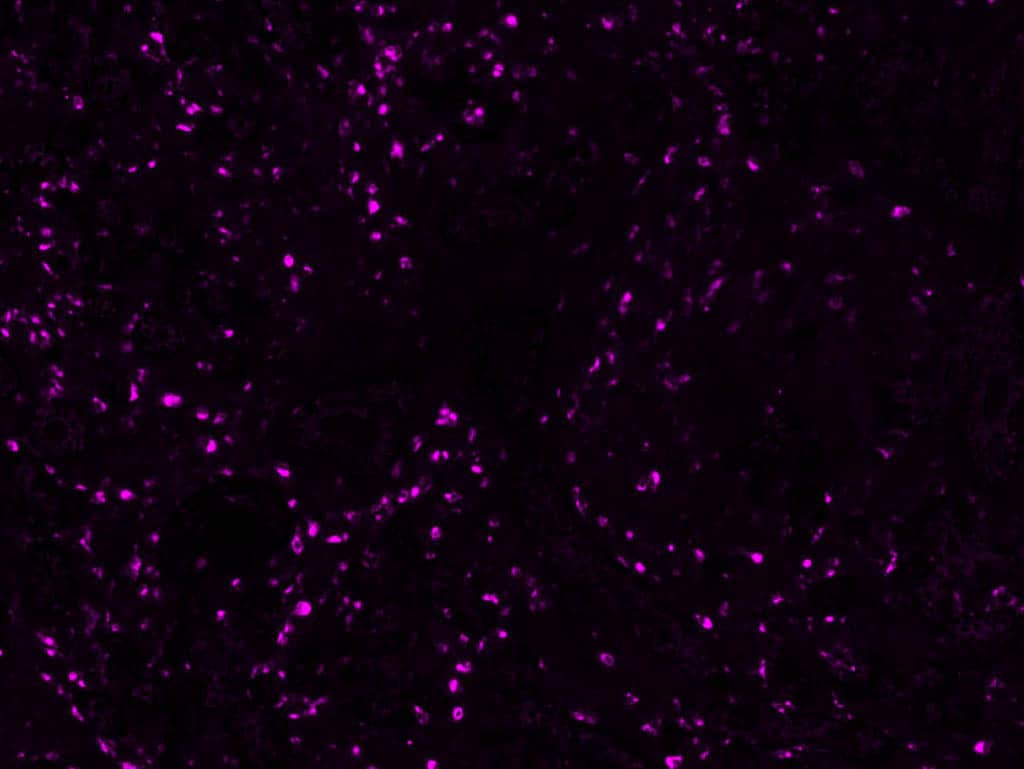
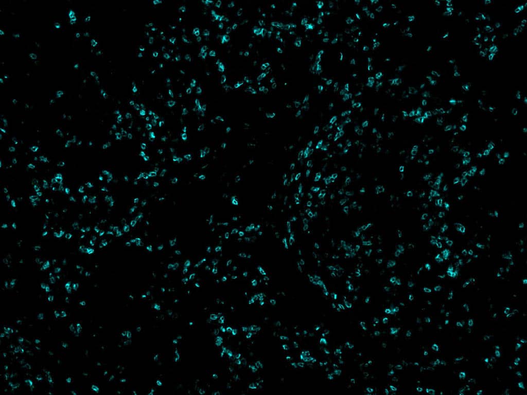
The images below include PD1, FoxP3, PDL1, PanCK, CD68 and CD8. They combine to form the image above allowing visualization of 6 biomarkers on one slide.
Reach out to get the latest in pricing and services.
© All rights reserved
Please fill out the form below to access the content.
Please fill out the form below to access the content.
Please fill out the form below to access the content.